
Identifying the Stages of Meiosis
- Cells undergoing meiosis can be observed and photographed using specialised microscopes
- The different stages of meiosis have distinctive characteristics meaning they can be identified from photomicrographs or diagrams
Meiosis I or Meiosis II
- Homologous chromosomes pair up side by side in meiosis I only
- This means if there are pairs of chromosomes in a diagram or photomicrograph meiosis I must be occurring
- The number of cells forming can help distinguish between meiosis I and II
- If there are two new cells forming it is meiosis I but if there are four new cells forming it is meiosis II
The distinguishing features at each stage of Meiosis I
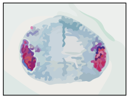
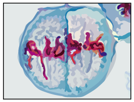
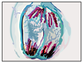
- Prophase I: Homologous pairs of chromosomes are visible
- Metaphase I: Homologous pairs are lined up side by side along the equator of spindle
- Anaphase I: Whole chromosomes are being pulled to opposite poles with centromeres intact
- Telophase I: There are 2 groups of condensed chromosomes around which nuclei membranes are forming
- Cytokinesis: Cytoplasm is dividing and cell membrane is pinching inwards to form two cells
The distinguishing features at each stage of Meiosis II
- Prophase II: Single whole chromosomes are visible
- Metaphase II: Single whole chromosomes are lined up along the equator of the spindle in single file (at 90 degree angle to the old spindle)
- Anaphase II: Centromeres divide and chromatids are being pulled to opposite poles
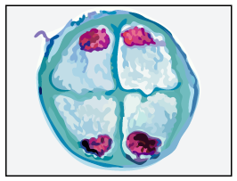
- Telophase II: Nuclei are forming around the 4 groups of condensed chromosomes
- Cytokinesis: Cytoplasm is dividing and four haploid cells are forming
Identifying the stages of meiosis table
| Stage | Micrograph |
|
Prophase I |
 |
|
Metaphase I |
 |
| Anaphase I Whole chromosomes are being pulled away from the middle |
 |
| Telophase I There are two groups of chromosomes at each pole The nucleus is reforming and the cytoplasm is pinching in |
 |
| Prophase II Two groups of chromosomes are visible as the DNA condenses |
 |
| Metaphase II Chromosomes line up along the middle of the spindles in single-file |
 |
| Anaphase II Chromatids are pulled away from the middle of the spindles |
 |
| Telophase II There are four groups of chromosomes and the cytoplasm is pinching in |
 |
Meiosis I Photomicrographs

Prophase I, Metaphase I , Anaphase I and Telophase I as seen in photomicrographs
Meiosis II Photomicrographs

Prophase II, Metaphase II , Anaphase II and Telophase II as seen in photomicrographs
Exam Tip
The acronym PMAT can help you remember what is happening in each stage:
- P for Prophase where chromosomes are Preparing to divide
- M for Metaphase for the middle of the spindle and cell which is where the chromosomes will be lined up.
- A for Anaphase, remember A for away from the middle to the poles, which is where the chromosomes / chromatids are being pulled
- T for telophase where we have Two cells (for meiosis I at least!)
