Calculation of Cardiac Output (Edexcel International A Level (IAL) Biology): Revision Note
Exam code: YBI11

Calculation of Cardiac Output
Cardiac output (CO) is the term used to describe the volume of blood that is pumped by the heart (the left and right ventricle) per unit of time
An average adult has a cardiac output of roughly 4.7 litres of blood per minute when at rest
Individuals who are fitter often have higher cardiac outputs due to having thicker and stronger ventricular muscles in their hearts
Cardiac output increases when an individual is exercising
This is so that the blood supply can match the increased metabolic demands of the cells
The CO of an individual can be calculated using their heart rate and stroke volume
Heart rate is the number of times a heart beats per minute
This can also be described as the number of cardiac cycles per minute
Stroke volume is the volume of blood pumped out of the left ventricle during one cardiac cycle

Graph showing the changes in the volume of the left ventricle within one cardiac cycle; this is the stroke volume
Calculating cardiac output
Cardiac output is found by multiplying the heart rate by the stroke volume:
Cardiac output = heart rate x stroke volume
The equation can be rearranged to find the heart rate and stroke volume if required:
Heart rate = cardiac output ÷ stroke volume
Stroke volume = cardiac output ÷ heart rate
Cardiac output is measured in cm3 min-1
Heart rate is measured in beats per min (bpm)
Stroke volume is measured in cm3
Worked Example
A woman took 0.833 seconds to complete a single cardiac cycle. The stroke volume of her heart was measured at 75 cm3. Calculate the cardiac output. Give your answer in dm3.
Answer:
Step 1: Find the heart rate
One cardiac cycle (atrial systole, ventricular systole and diastole) takes 0.833 seconds
To find the number of cardiac cycles completed in a minute, divide 60 (seconds) by the time taken for one cycle, 0.833 seconds
Heart rate = 60 ÷ 0.833
= 72 bpm
Step 2: Insert relevant figures into the equation
Cardiac output = heart rate x stroke volume
Cardiac output = 72 x 75 = 5 400 cm3
Step 3: Convert to dm3
1 000 cm3 = 1 dm3
Cardiac output = 5 400 ÷ 1 000 =
5.40 dm3
Worked Example
An athlete runs a 10 km race, after which his heart rate was measured at 110 bpm and his cardiac output was determined to be 9,800 cm3.
Calculate the stroke volume after the race.
Answer:
Stroke volume = cardiac output ÷ heart rate
Stroke volume = 9,800 ÷ 110
Stroke volume = 89.1 cm3
Examiner Tips and Tricks
1 dm3 is equal to 1000 cm3. It can be useful to convert all the figures found in the first question into the same units before starting your working out, that way you are less likely to make any mistakes!
Effects of Variability of Cardiac Output
During exercise, muscle contraction occurs more frequently, requiring more energy
The rate of aerobic respiration increases to meet the increase in energy demand
This means that cells require more oxygen to be delivered to them, while producing more carbon dioxide as a waste product of respiration
The body will accommodate this by making the following changes:
Increase the rate and depth of breathing which will increase the amount of oxygen entering the lungs and bloodstream, while getting rid of more carbon dioxide
Increase the heart rate which will transport the oxygen (and glucose) to the muscles much faster, while removing the additional carbon dioxide produced due to the increased rate of respiration
Control of the breathing rate
Breathing rate is controlled by the ventilation centres (also called respiratory centres) in the medulla oblongata
This is one of the three regions that make up the brainstem, it transfers nerve messages from the brain to the spinal cord
The inspiratory centre controls the movement of air into the lungs (inhalation)
The expiratory centre controls the movement of air out of the lungs (exhalation)
The inspiratory centre in the medulla oblongata has the following effect on breathing:
It sends nerve impulses along motor neurons to the intercostal muscles of the ribs and diaphragm muscles
These muscles will contract and cause the volume of the chest to increase
This lowers the air pressure in the lungs to slightly below atmospheric pressure
An impulse is also sent to the expiratory centre to inhibit its action
Due to the difference in pressure between the lungs and outside air, air will flow into the lungs
Stretch receptors in the lungs are stimulated as they inflate with air
Nerve impulses are sent back to the medulla oblongata which will inhibit the inspiratory centre
The expiratory centre is no longer inhibited and will bring about the following changes:
It sends nerve impulses to the intercostal and diaphragm muscles
These muscles will relax and cause the volume of the chest to decrease
This increases the air pressure in the lungs to slightly above atmospheric pressure
Due to the higher pressure in the lungs, air will flow out of the lungs
As the lungs deflate, the stretch receptors become inactive which means that the inspiratory centre is no longer inhibited and the next breathing cycle can begin

The process of breathing in (inhalation)

The process of breathing out (exhalation)
Effect of exercise
The extra carbon dioxide that is produced due to the increase in the rate of respiration during exercise dissolves in the blood to form carbonic acid
This quickly dissociates into hydrogen ions (H+) and hydrogencarbonate ions (HCO3-)
The increase in the concentration of H+ ions will decrease the pH of the blood (it becomes more acidic)
The decrease in pH is detected by receptors sensitive to changes in the chemical composition of blood
These are called chemoreceptors and they are located in several places
In the ventilation centre of the medulla oblongata
They are also present as clusters of cells in the aorta (aortic bodies) and the carotid arteries (carotid bodies)
Once they are stimulated a nerve impulse is sent to the medulla oblongata
The medulla oblongata will then send more frequent nerve impulses to the intercostal and diaphragm muscles to increase the rate and strength of contractions
This increases the breathing rate and depth
This results in more oxygen entering the lungs (and bloodstream), while more carbon dioxide can be exhaled and thus be removed from the bloodstream
The decrease in carbon dioxide levels will result in the blood pH returning back to normal, which leads to the breathing rate returning to normal
The volume of air that moves in and out of the lungs during a set time period (e.g. a minute) is known as the ventilation rate
The ventilation rate increases during exercise due to the increase in breathing rate and depth
Control of the heart rate
The cardiovascular control centre in the medulla oblongata unconsciously controls the heart rate by controlling the rate at which the sinoatrial node (SAN) generates electrical impulses
These electrical impulses cause the atria to contract and therefore determines the rhythm of a heartbeat
Changes in the internal environment of the body (e.g. blood pressure, oxygen levels) can result in a change in the heart rate
These changes act as stimuli which is detected by baroreceptors and chemoreceptors
Baroreceptors are found in the aortic and carotid bodies and they are stimulated by high and low blood pressure
Chemoreceptors are found in the medulla oblongata, as well as in the aortic and carotid bodies
They are stimulated by changes in the levels of carbon dioxide and oxygen in the blood, as well as blood pH
Once stimulated, these receptors will send electrical impulses to the medulla oblongata
The cardiovascular control centre in the medulla oblongata will respond by sending impulses to the SAN along sympathetic or parasympathetic neurones
Each of these neurones release different neurotransmitters which will affect the SAN in a different way
Sympathetic neurones will increase the rate at which the SAN generates electrical impulses, thus speeding up the heart rate
These neurones form part of the sympathetic nervous system which prepares the body for action ('fight or flight' response) and increases the heart rate during exercise
Parasympathetic neurones will decrease the rate at which the SAN fires, thus slowing down the heart rate
These neurones form part of the parasympathetic nervous system which calms the body down after action ('rest and digest' response) and decreases the heart rate after exercise
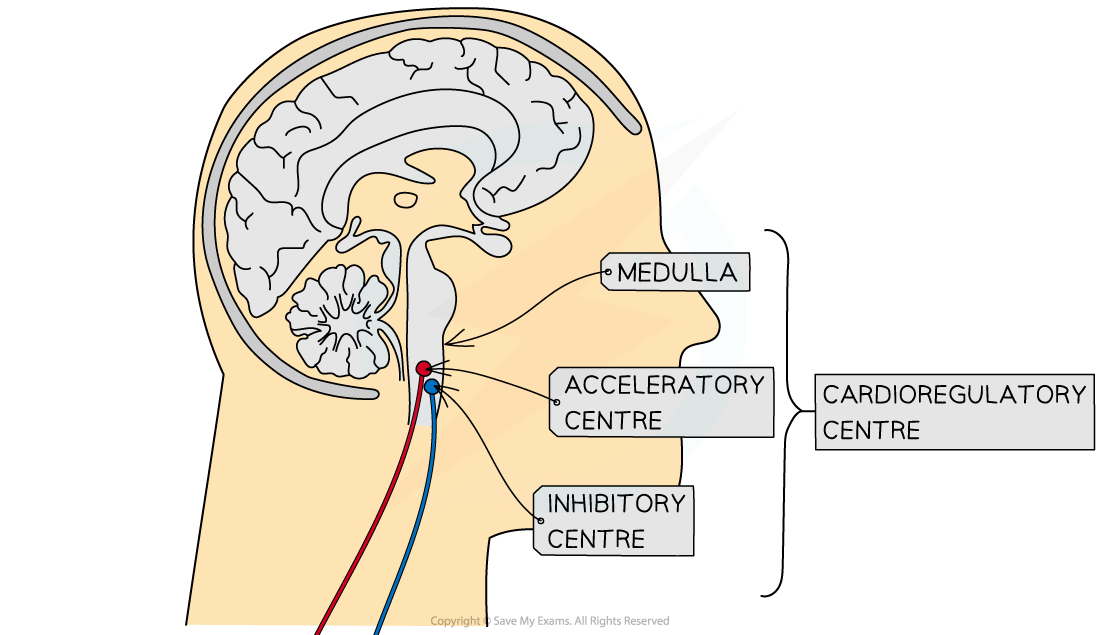
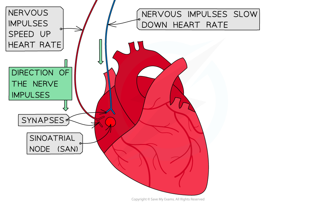
Nervous control of the heart rate by the cardioregulatory centre (also known as the cardiovascular control centre). Sympathetic neurones (indicated in red) will speed up the heart rate while parasympathetic neurones (indicated in blue) will slow the heart rate down
Changes in heart rate
The heart will respond in different way depending on the stimulus that it receives
High blood pressure
Detected by baroreceptors which send impulses to cardiovascular control centre
It sends impulses along parasympathetic neurones which secrete the neurotransmitter acetylcholine
Acetylcholine binds to receptors on SAN causing it to fire less frequently
Heart rate slows down and blood pressure decreases back to normal
Low blood pressure
Detected by baroreceptors which send impulses to cardiovascular control centre
It sends impulses along sympathetic neurones which secrete the neurotransmitter noradrenaline
Noradrenaline binds to receptors on SAN causing it to fire more frequently
Heart rate speeds up and blood pressure increases back to normal
High blood O2 / Low CO2 / high pH levels
Detected by chemoreceptors which send impulses to cardiovascular control centre
It sends impulses along parasympathetic neurones which secrete the neurotransmitter acetylcholine
Acetylcholine binds to receptors on SAN causing it to fire less frequently
Heart rate slows down and O2 / CO2 and pH levels return to normal
Low blood O2 / High CO2 / low pH levels (during exercise)
Detected by chemoreceptors which send impulses to cardiovascular control centre
It sends impulses along sympathetic neurones which secrete the neurotransmitter noradrenaline
Noradrenaline binds to receptors on SAN causing it to fire more frequently
Heart rate speeds up and O2 / CO2 and pH levels return to normal

You've read 0 of your 5 free revision notes this week
Unlock more, it's free!
Did this page help you?