Maintaining Water & Nitrogen Balance in the Body (AQA GCSE Biology): Revision Note
Exam code: 8461

Water loss in the body
Maintaining water levels in the body is vital to prevent harmful changes occurring to cells of the body as a result of osmosis
The cytoplasm of all cells is largely composed of water, as is the blood plasma
If body cells lose or gain too much water by osmosis they do not function efficiently:
Too much water in the blood results in cells swelling as water moves into them; this has a diluting effect and can lead to cell lysis (bursting)
Too little water in the blood (or too high an ion concentration) and the cells lose water by osmosis; this has a dehydrating effect and can lead to cell death
There are two sources of water in the body:
water produced as a result of aerobic respiration
water in the diet
Water is lost from the body in the following ways:
Water leaves the body via the lungs during exhalation
Water, ions and urea are lost from the skin in sweat
The lungs and skin have no control over how much water, ions or urea is lost
Loss of excess water, ions and urea is controlled by the kidneys when they filter the blood to produce urine

Deamination
Higher Tier Only
The digestion of proteins from the diet results in excess amino acids which need to be excreted safely
They cannot be stored by the body
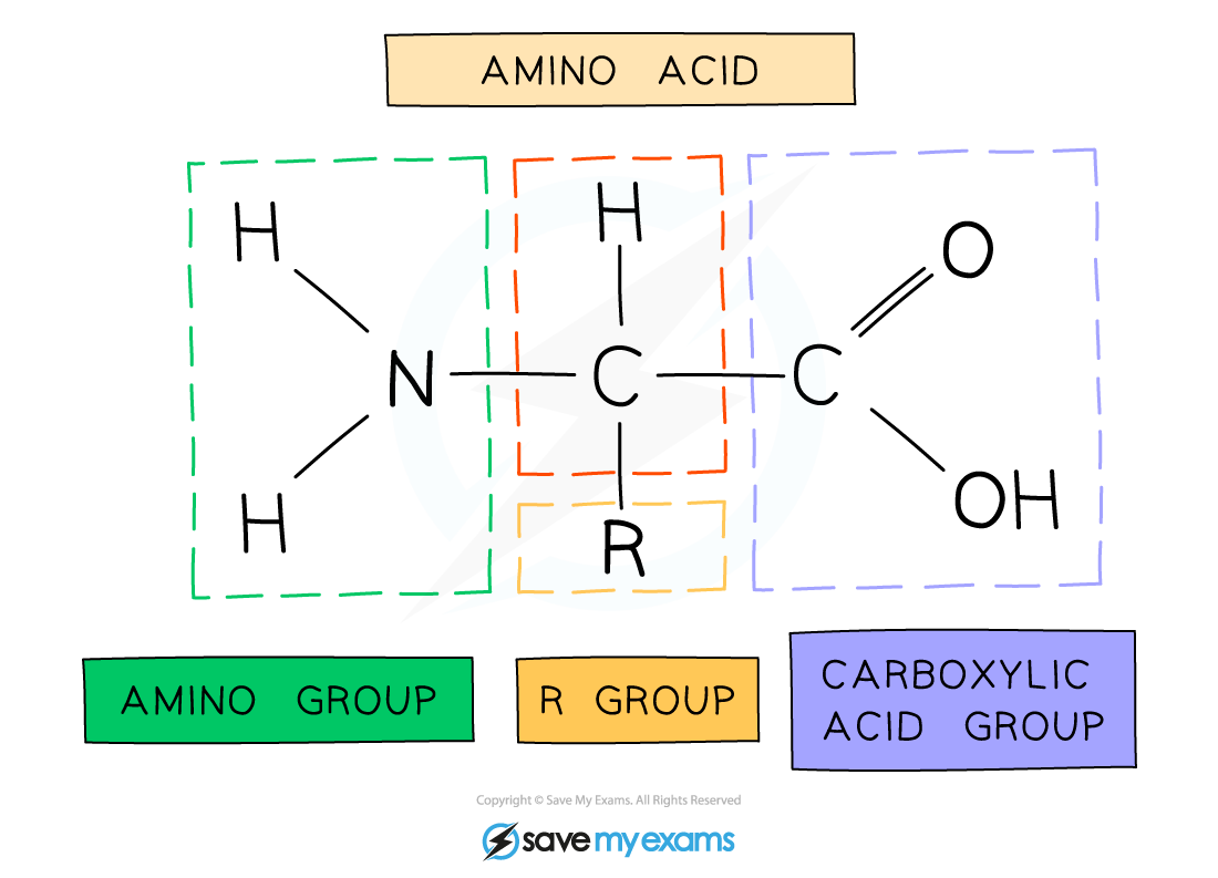
Excess amino acids are process in the liver in a process known as deamination
Deamination removes the amino group; the amino acid is said to be deaminated
Enzymes in the liver split up amino acid molecules and ammonia is formed
Ammonia is toxic to cells and so it is immediately converted to urea which is excreted by the kidneys
Structure & function of the kidney
The kidneys help to control the water content of the body and the concentrations of substances (such as mineral ions) dissolved in the fluids of the body
Kidneys filter the blood as follows:
Blood flows into the kidneys at high pressure
Small molecules are filtered out of the the blood into the kidney filtrate; these include glucose, urea, water and ions
Large molecules, such as proteins, are too large to pass through the filtration mechanism and remain in the blood
The kidneys selectively reabsorb substances needed by the body
In a healthy kidney, this includes:
all of the glucose
some ions
some water
Anything not reabsorbed forms urine, which is stored in the bladder until it is excreted
Urea is not selectively reabsorbed and so is excreted in the urine

Examiner Tips and Tricks
Be sure to use technical terminology when describing the events in the kidney, for example substances are 'reabsorbed' rather than just 'absorbed'.
Interpreting information about kidney function
Examiner Tips and Tricks
You should be able to translate tables and bar charts of glucose, ions and urea before and after filtration – expect to see this in an exam.
Control of water levels
Higher Tier Only
The volume of water lost in urine is controlled in the kidneys
This is achieved by regulating the volume of water that is reabsorbed by the kidney tubules before urine passes into the bladder
Kidney tubules are tiny tubes through which the filtrate flows inside the kidney
When blood water level is low:
low blood water content is detected by the pituitary gland
the pituitary gland releases the hormone ADH
ADH travels in the blood to the kidneys where it increases the permeability of the kidney tubules to water
more water is reabsorbed into the blood
a smaller volume of concentrated urine is produced
When blood water level is high:
high blood water content is detected by the pituitary gland
the pituitary gland releases less ADH
the permeability of the kidney tubules to water decreases
less water is reabsorbed into the blood
a higher volume of dilute urine is produced
This is an example of negative feedback control

Kidney failure
The kidneys might not work properly for several reasons, including accidents or disease
Humans can survive with one functioning kidney, but if both are damaged then there will quickly be a build-up of toxic wastes in the body which will be fatal if not removed
Kidney failure can be treated by:
dialysis
kidney transplant
Dialysis
Patients are connected to a dialysis machine which acts as an artificial kidney to remove most of the urea and restore/maintain the water and salt balance of the blood
Unfiltered blood is taken from an artery in the arm, and pumped into the dialysis machine
Inside the machine the blood and dialysis fluid are separated by a partially permeable membrane
Dialysis fluid contains:
A glucose concentration similar to a normal level in blood
A concentration of salts similar to a normal level in blood
No urea
Exchange of substances occurs between the blood and the fluid
This exchange relies on concentration gradients

Kidney transplants
Kidney transplants involve transferring a healthy kidney from a donor into the body of a recipient

If a healthy, close matched kidney is available, then the benefits of a transplant over dialysis include:
The patient has much more freedom as they are not tied to having dialysis several times a week in one place
Their diets can be much less restrictive than they are when on dialysis
Use of dialysis machines is very expensive and so this cost is removed
A kidney transplant is a long term solution whereas dialysis will only work for a limited time
However, there are several disadvantages to kidney transplants, including:
Donors won’t have the same antigens on cell surfaces so there will be some immune response to the new kidney
risk of rejection is reduced—but not removed—by ‘tissue typing’ the donor and the recipient first
Recipients must take immunosuppressant drugs for the rest of their lives, which can have long term side effects and leave the patient vulnerable to infections
There are not enough donors to cope with demand
Examiner Tips and Tricks
When answering questions about dialysis, the best answers will refer to differences in concentration gradients between the dialysis fluid and the blood, and use this to explain why substances move in certain directions.

Unlock more, it's free!
Join the 100,000+ Students that ❤️ Save My Exams
the (exam) results speak for themselves:

Was this revision note helpful?
Build on this topic