Syllabus Edition

First teaching 2023

First exams 2025

|

The Heart (CIE AS Biology)

Exam Questions

3 hours45 questions
11 mark

What is systolic blood pressure?

  • The blood pressure in the arteries when the heart is relaxing.

  • The maximum blood pressure in the right ventricle.

  • The blood pressure in the left ventricle at the end of a contraction.

  • The maximum blood pressure in the arteries.

Did this page help you?

21 mark

Which structure is correctly labelled on the diagram below?

q2-cie-8-3

  • Pulmonary vein

  • Pulmonary artery

  • Aorta

  • Vena cava

Did this page help you?

31 mark

The graph shows the pressure in different parts of the heart during one cardiac cycle

q3-cie-8-3

At 0.2 seconds which part of the heart is responding to the excitatory stimulus?

q3-cie-8-3-2

    Did this page help you?

    41 mark

    During ventricular diastole what happens in the heart?

    • The pressure in the left atrium rises more than the pressure in the right atrium.

    • The pressure in the atria rises above the pressure in the ventricles.

    • The atrioventricular valves open.

    • All semilunar valves open.

    Did this page help you?

    51 mark

    The following are some features of blood vessels. 

    Which feature enables the aorta to withstand the high pressure at ventricular systole?

    • Smooth muscle and small lumen

    • Elastin fibres and large lumen

    • Collagen fibres and endometrium

    • Collagen fibres and elastin fibres

    Did this page help you?

    61 mark

    What happens in the left ventricle during a contraction?

    • The pressure in the left ventricle becomes greater than the pressure in the aorta.

    • The bicuspid valve closes and semilunar valves in the aorta close.

    • The bicuspid valve opens and the semilunar valve in the aorta opens.

    • The pressure in the left atrium becomes greater than the pressure in the left ventricle.

    Did this page help you?

    71 mark

    When the pressure in the ventricles in the heart is at its maximum what is the state of the valves?

      semilunar valves atrioventricular valves
    A open open
    B open closed
    C closed open
    D closed closed

    Did this page help you?

    81 mark

    The characteristic ‘lub-dub’ sound of the heartbeat is caused by the movement of valves in the heart.

    What causes the first sound after atrial systole in the cardiac cycle?

    1 closing of the semilunar valves
    2 opening of the semilunar valves
    3 closing of the atrioventricular valves

    • 1 and 3

    • 1 and 2

    • 1 only

    • 3 only

    Did this page help you?

    91 mark

    Which of the following statements happens during ventricular systole in a human heartbeat?

    1

    ventricular pressure increases

    2

    atrial pressure does not change

    3

    aortic pressure increases

    • 1 and 2

    • 1 and 3

    • 2 and 3

    • 3 only

    Did this page help you?

    101 mark

    Purkyne (Purkinje) tissue is found in the mammalian heart. What is its function?

    • To separate oxygenated blood from deoxygenated blood.

    • To reduce the spontaneous contraction rate of the heart.

    • To conduct a wave of electrical excitation over the ventricles.

    • To conduct a wave of electrical excitation over the atria.

    Did this page help you?

    11 mark

    The graph shows the pressure in different parts of the heart during one cardiac cycle

    q11-cie-8-3

    At which point does the semilunar valve of the aorta close?

      Did this page help you?

      21 mark

      The diagram below shows the heart and associated blood vessels.

      q12-cie-8-3

      Which of the following would be correct for the flow of blood through the heart from the body?

      • 4 → 3 → 2 → 1

      • 3 → 4 → 2 → 1

      • 2 → 1 → 3 → 4

      • 1 → 2 → 3 → 4

      Did this page help you?

      31 mark

      The graph below shows the pressure in different parts of the left side of the heart during one cardiac cycle.

      At the end of which section in the graph (A, B, C or D) would the ventricle be full of blood?

      q13-cie-8-3

        Did this page help you?

        41 mark

        The muscle of the left-hand side of the heart is thicker than the right-hand side. 

        Which of the following are consequences of this difference in muscle thickness?

        1. The left side of the heart generates higher blood pressure
        2. Blood leaving the right ventricle travels a greater distance than blood leaving the left ventricle
        3. The left ventricle delivers blood to the body
        4. The right ventricle generates lower pressure than the left-hand side
        • 1, 2 and 3

        • 2, 3 and 4

        • 1, 3 and 4

        • 3 only

        Did this page help you?

        51 mark

        The narrowing of the aortic semilunar valve is a heart disorder called aortic stenosis.

        Which of the following would describe the effect of aortic stenosis on the heart structure and function?

        • The wall of the left ventricle thickens, leading to an enlarged heart and an inability to relax and fill during diastole.

        • There is less cardiac muscle in the left ventricle and reduced diastolic blood pressure, caused by the smaller blood volume entering the left atrium.

        • The cardiac muscle of the left ventricle wall is thinned by blood leaking out of the left ventricle during ventricular diastole.

        • The tendons of the heart valves are weakened by blood being forced back through the bicuspid/left atrioventricular valve into the left atrium.

        Did this page help you?

        6
        Sme Calculator
        1 mark

        The diagram below shows two cardiac cycles of a patient. The events of the cycle are placed next to a timescale.

        q16-cie-8-3

        How fast is the patient’s heart beating?

        • 90

        • 80

        • 75

        • 72

        Did this page help you?

        71 mark

        The electrical excitation of the ventricles of the heart are thought to be improved by taking fish oils.

        What part of the heart could the fish oils help?

        • Vagus nerve

        • Sinoatrial node (SAN)

        • Atrioventricular node (AVN)

        • Purkyne tissue

        Did this page help you?

        81 mark

        The following parts of the heart all control the heartbeat.

        1

        Purkyne tissue

        2

        atrioventricular node (AVN)

        3

        sinoatrial node (SAN)

        Which row in the table below is correct for ventricular contraction and atrial contraction?


        Ventricular contraction

        Atrial contraction

        A

        Purkyne tissue carries a wave of excitation

        SAN produces a wave of excitation

        B

        Purkyne tissue carries a wave of excitation

        SAN and AVN produce a wave of excitation

        C

        AVN produces a wave of excitation

        Purkyne tissue carries a wave of excitation

        D

        SAN produces a wave of excitation

        AVN produces a wave of excitation

          Did this page help you?

          91 mark

          The diagram shows a fault in the wall of the atria.

          q19-cie-8-3-

          Which of the following would describe the effect of this fault?

          • Irregular heartbeat

          • Ventricular systole is delayed

          • Increased pressure in the pulmonary artery

          • Reduced oxygen saturation of haemoglobin

          Did this page help you?

          101 mark

          The table shows some changing conditions in the body.

          Which row would cause the largest rise in cardiac output?

            aortic blood pressure blood pressure in the vena cava carbon dioxide concentration of the blood frequency of impulses in the Vagus nerve

          A

          B

          C

          D

          no change

          no change

          decrease

          decrease

          decrease

          decrease

          no change

          no change

          ncrease

          no change

          increase

          no change

          no change

          increase

          no change

          increase

            Did this page help you?

            11 mark

            The volume of blood pumped by each contraction of the ventricle is called the stroke volume. The volume of blood in the ventricle before systole is called the ventricular end-diastolic volume.

            The following graphs show the relationship between stroke volume and ventricular
            end-diastolic volume.

            Which one is correct?

            q21-cie-83

            q21-cie-8-3-2                       

              Did this page help you?

              21 mark

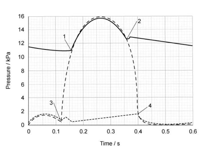
              The graph shows the pressure changes in the left side of the heart over time. The cardiac cycle shown lasts 0.6 seconds (s). 

              The points on the graph 1, 2, 3 and 4 show when the atrioventricular and semilunar valves are open or closed.

              q22-cie-8-3

              What is the total time in one cardiac cycle when the atrioventricular valves and the semilunar valves are both closed at the same time?

              • 0.21 s

              • 0.08 s

              • 0.04 s

              • 0.03 s

              Did this page help you?

              31 mark

              The graph shows the pressure changes during a cardiac cycle 

              screenshot-2023-05-13-235131

              Which row correctly identifies Q, R, S and T?


              Q

              R

              S

              T

              A

              semilunar valves close

              atrioventricular valves open

              atrioventricular valves close

              semilunar valves open

              B

              semilunar valves open

              atrioventricular valves close

              atrioventricular valves open

              semilunar valves close

              C

              atrioventricular valves close

              semilunar valves close

              semilunar valves open

              atrioventricular valves open

              D

              atrioventricular valves close

              semilunar valves open

              semilunar valves close

              atrioventricular valves open

                Did this page help you?

                41 mark

                The diagram shows a horizontal cross section of a human heart. The atria have been removed so the valves can be seen.

                screenshot-2023-05-13-235953

                 Which of the labels shows the valve that is pushed open when oxygenated blood is entering a ventricle?

                  Did this page help you?

                  5
                  Sme Calculator
                  1 mark

                  The graph below shows the blood pressure changes in the aorta and left ventricle during two complete cardiac cycles.

                  cie-ial-8-3-h-q5

                  Fig. 1

                  What is this person's heart rate?

                  • 62.5 bpm

                  • 60 bpm

                  • 54.5 bpm

                  • 31.3 bpm

                  Did this page help you?

                  61 mark

                  The table below describes events during the cardiac cycle. 

                  Which row is correct?


                  nerve impulses from atrioventricular node (AVN) to

                  nerve impulses from Purkyne tissue (PT) to

                  nerve impulses from the sinoatrial node (SAN) to

                  A

                  PT

                  the atria

                  AVN

                  B

                  SAN

                  the atria

                  PT

                  C

                  PT

                  the ventricles

                  AVN

                  D

                  SAN

                  the ventricles

                  PT

                    Did this page help you?

                    71 mark

                    After entering the right side of the heart, a red blood cell will pass through or pass by the following structures:

                    1

                    tricuspid valve

                    2

                    semilunar valve

                    3

                    right atrium

                    4

                    right ventricle

                    5

                    sinoatrial node

                    Which option shows the correct order that red blood cells will pass through?

                    • 4 → 3 → 5 → 2 → 1

                    • 5 → 3 → 1 → 4 → 2

                    • 2 → 1 → 5 → 3 → 4

                    • 5 → 1 → 2 → 3 → 4

                    Did this page help you?

                    81 mark

                    A disease that causes a lower than normal heart rate is called ‘heart block’. When a doctor was treating a patient with this condition, he found that they had normal electrical impulses but these were not correctly conducted to the ventricles. 

                    Which of the following could be faulty in the patient?

                    1

                    atrioventricular node (AVN)

                    2

                    sinoatrial node (SAN)

                    3

                    Purkyne tissue

                    • 1 and 2

                    • 1 and 3

                    • 2 and 3

                    • 3 only

                    Did this page help you?

                    91 mark

                    The diagram is of the external heart structure and blood vessels.

                    screenshot-2023-05-14-001410

                    Which row of the table below shows the combination of blood vessels that supply blood to the heart and carry blood away from the heart?

                      To the heart Away from the heart
                    A 2 and 3 1 and 4
                    B 4 and 1 2 and 3
                    C 3 and 4 1 and 2
                    D 1 and 2 3 and 4

                      Did this page help you?

                      101 mark

                      The graph shows the pressure changes in the left side of the heart over time.

                      screenshot-2023-05-14-001726

                      Which row of the table below shows what is happening at points 1, 2, 3 and 4 to the semilunar valves and the atrioventricular valves?

                        Semilunar valve Atrioventricular valve
                        Opens Closes Opens Closes
                      A 2 3 1 4
                      B 2 3 4 1
                      C 1 2 3 4
                      D 1 2 4 3

                        Did this page help you?