Eukaryotic Cell Structures & Functions (Cambridge (CIE) AS Biology): Revision Note
Exam code: 9700
Eukaryotic cell structures & functions
Cells can be divided into two broad types; eukaryotic and prokaryotic cells
Eukaryotic cells have a more complex ultrastructure than prokaryotic cells
The term ultrastructure refers to the internal structure of cells
The cytoplasm of eukaryotic cells is divided up into membrane-bound compartments called organelles
Cell organelles
Cell surface membrane
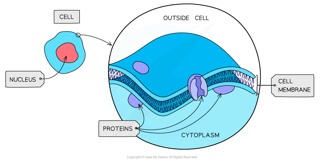
All cells are surrounded by a cell surface membrane which separates the inside of cells from their surroundings
Cell surface membranes controls the exchange of materials between the internal cell environment and the external environment
The membrane is described as being partially permeable as it allows the passage of some substances and not others
The cell membrane is formed from a phospholipid bilayer and spans a diameter of around 10 nm
Nucleus
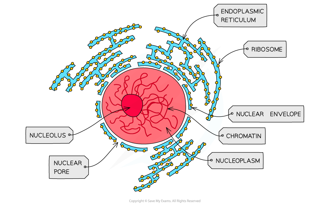
Present in all eukaryotic cells, the nucleus is a large organelle that is separated from the cytoplasm by a double membrane
The nucleus contains the DNA, which is arranged into chromosomes
Chromosomes contain DNA and proteins, which are collectively referred to as chromatin
The nuclear membrane is known as the nuclear envelope, and contains many pores
Nuclear pores are important channels for allowing mRNA and ribosomes to travel out of the nucleus, as well as allowing enzymes and signalling molecules to move in
The nucleus contains a region known as the nucleolus, which is the site of ribosome production
Rough and smooth endoplasmic reticulum
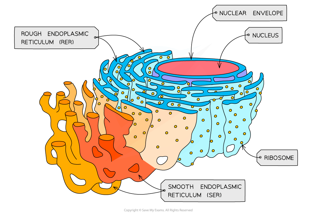
The endoplasmic reticulum (ER) is made up of a series of membranes that form flattened sacs within the cell cytoplasm
The ER is linked with the nuclear envelope
There are two distinct types of ER, with different roles within the cell
The rough endoplasmic reticulum (RER)
Continuous folds of membrane that are linked with the nuclear envelope
The surface of the RER is covered in ribosomes
The role of the RER is to process proteins that are produced on the ribosomes
The smooth endoplasmic reticulum (SER)
The SER does not have ribosomes on the surface
It is involved in the production of lipids, and of steroid hormones such as oestrogen and testosterone
Examiner Tips and Tricks
Be sure to always use the full name of the rough and smooth endoplasmic reticulum when you first refer these structures in an exam; marks are often not awarded for the abbreviations RER and SER in the absence of the full key terms.
Golgi body
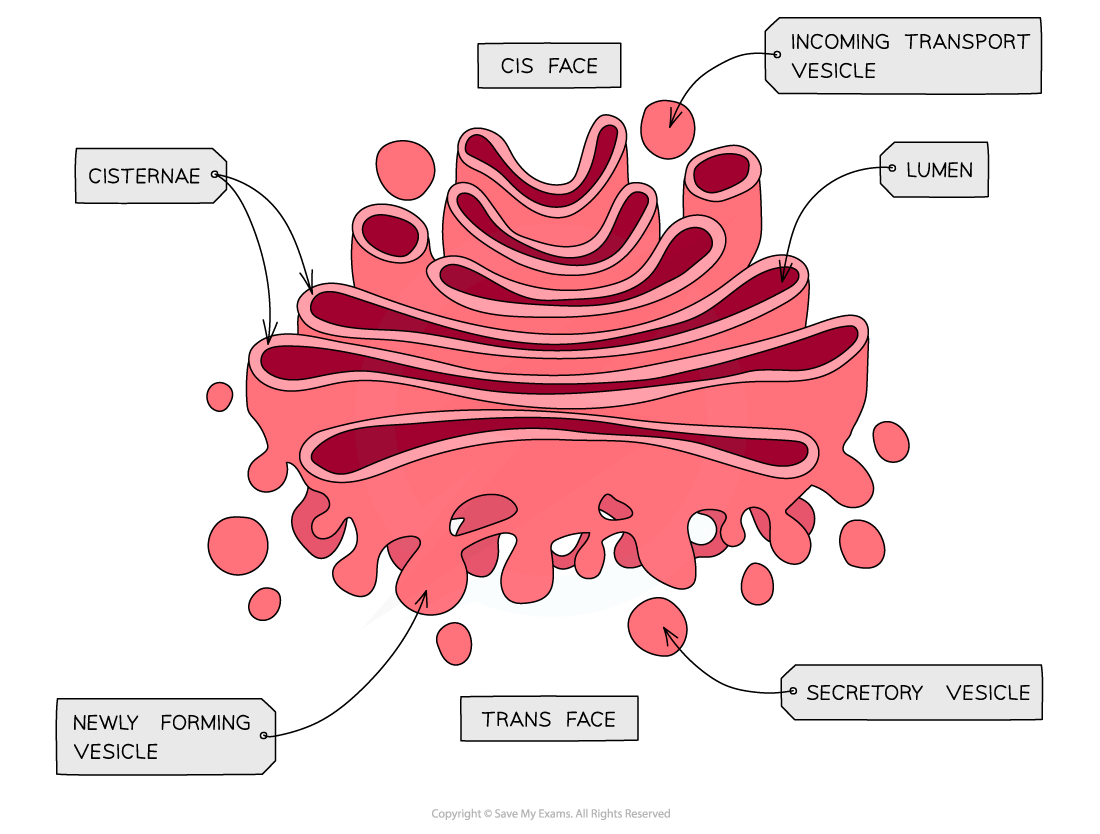
The Golgi body is often referred to as the Golgi apparatus or the Golgi complex
It consists of a series of flattened sacs of membrane
It can be clearly distinguished from the ER, as it is not connected to other membrane-bound compartments, and it has a distinctive 'wifi symbol' appearance
Its role is to modify proteins and package them into vesicles
Mitochondria
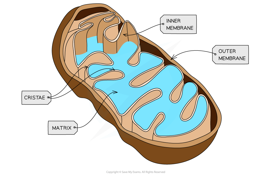
Mitochondria (singular mitochondrion) are relatively large organelles surrounded by a double-membrane
They are smaller than the nucleus and chloroplasts, but can be seen with a light microscope
The inner membrane is folded to form cristae
Mitochondria are the site of aerobic respiration within eukaryotic cells
The mitochondrial matrix contains enzymes needed for aerobic respiration
Small, circular pieces of DNA, known as mitochondrial DNA, and ribosomes are also found in the matrix
This allows the production of proteins required for respiration
Ribosomes
Ribosomes are found in the cytoplasm of all cells or as part of the rough endoplasmic reticulum in eukaryotic cells
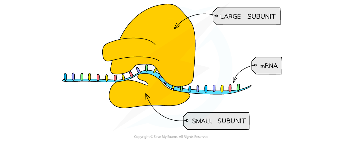
Each ribosome is a complex of ribosomal RNA (rRNA) and proteins
80S ribosomes (composed of 60S and 40S subunits) are found in eukaryotic cells
Smaller, 70S ribosomes (composed of 50S and 30S subunits) are found in prokaryotes, mitochondria and chloroplasts
Ribosomes are the site of translation during protein synthesis
Vesicles
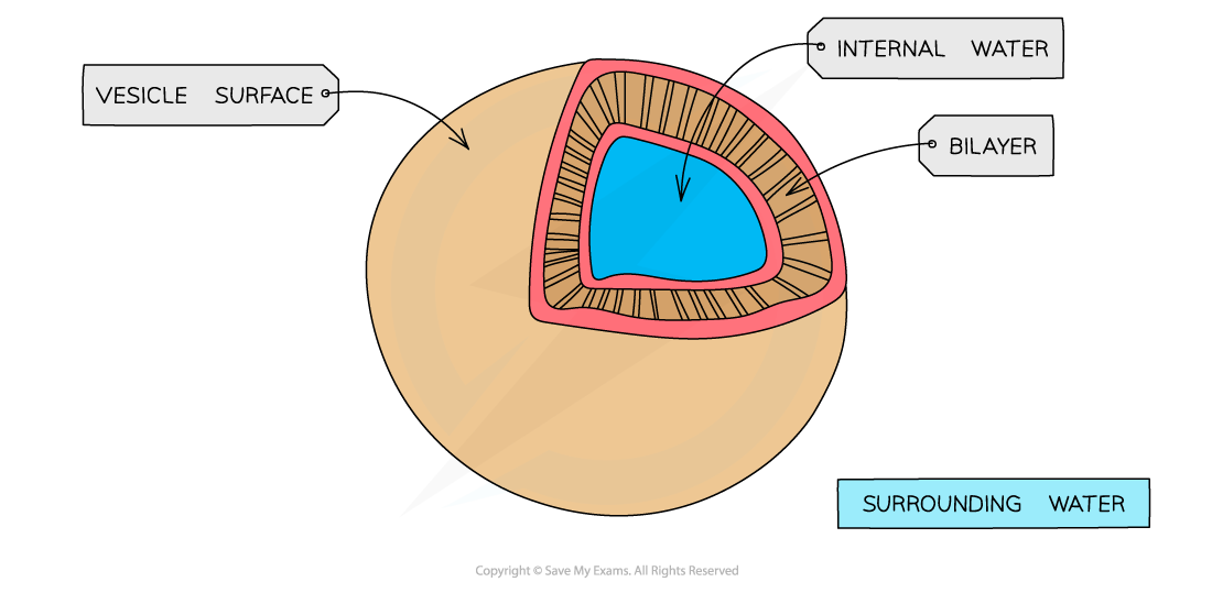
Vesicles are small, membrane-bound sacs used by cells for transport and storage
They can be pinched off the ends of the Golgi body; these are known as Golgi vesicles
They can fuse with the cell surface membrane to allow exocytosis, or bud from the membrane during endocytosis
Lysosomes
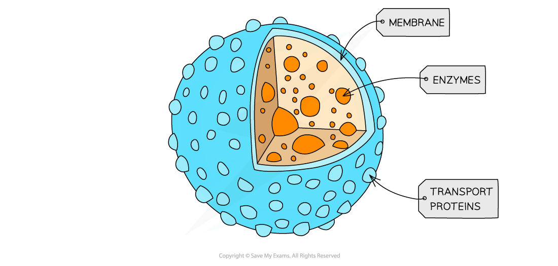
Lysosomes are specialised vesicles which contain hydrolytic enzymes
Hydrolytic enzymes break down biological molecules, e.g.
Waste materials, such as worn-out organelles
Engulfed pathogens during phagocytosis
Cell debris during apoptosis (programmed cell death)
Lysosome diagram
Centrioles
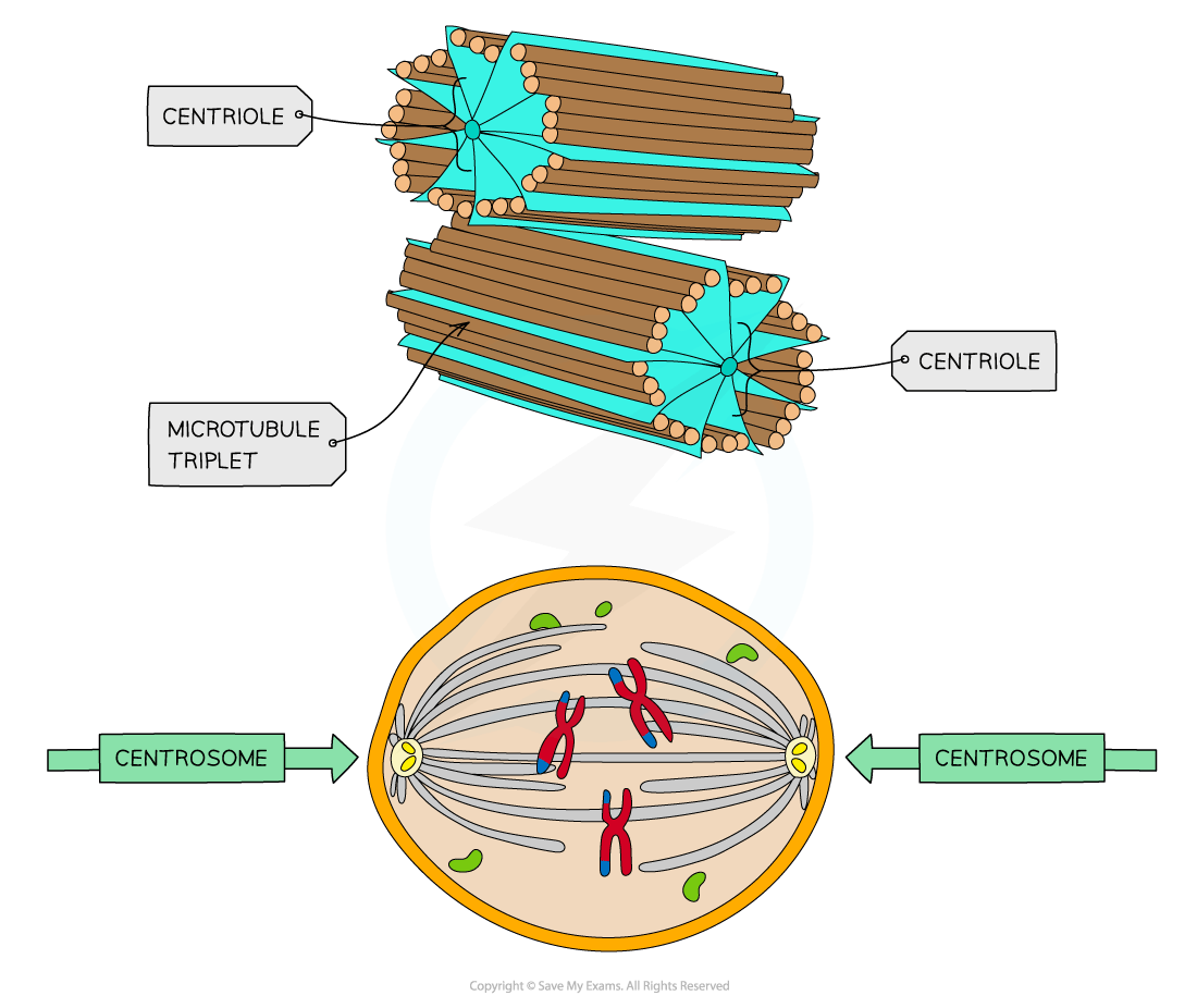
Centrioles are hollow fibres made of microtubules
Two centrioles at right angles to each other form a centrosome, which organises the spindle fibres during cell division
Note that centrioles are not found in flowering plants and fungi
Microtubules
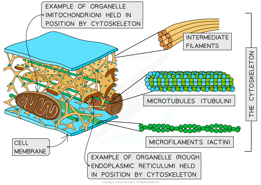
Microtubules are hollow tubes made of tubulin protein
α and β tubulin proteins combine to form dimers, which are then joined into protofilaments
Thirteen protofilaments in a cylinder make a microtubule
Microtubules make up the cytoskeleton of the cell
The cytoskeleton is used to provide support and movement of the cell

Cilia
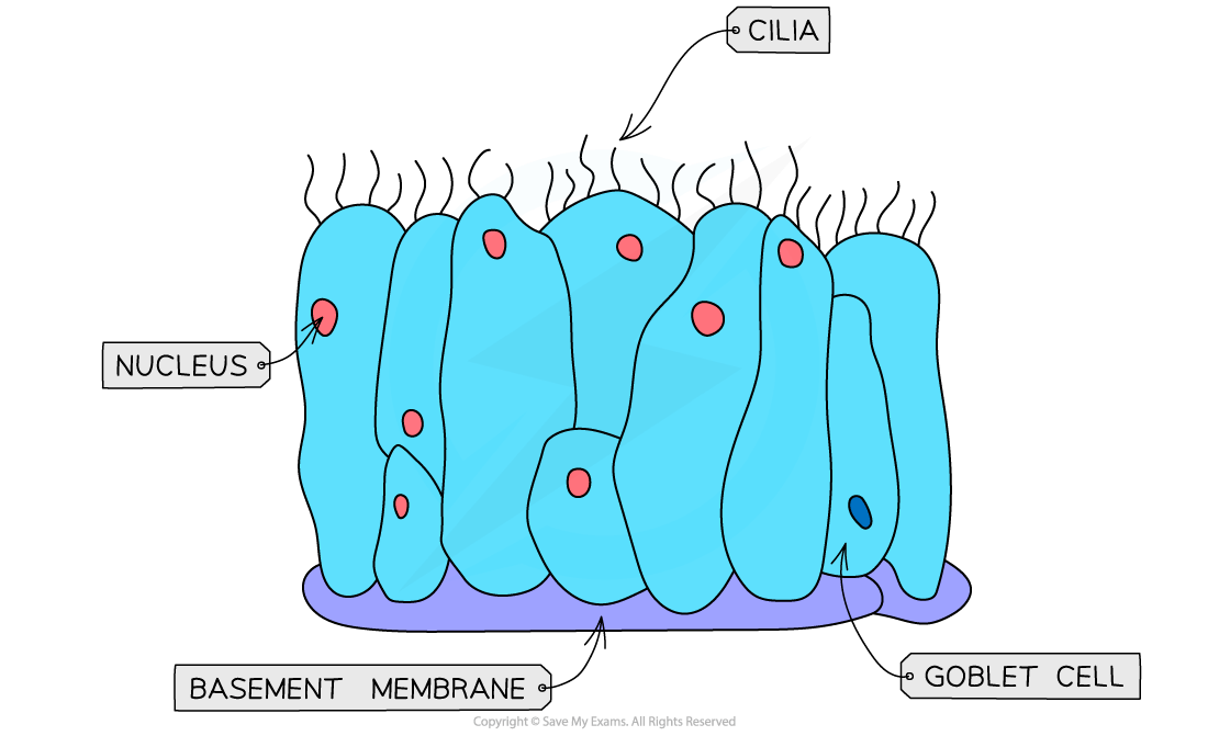
Cilia are hair-like projections made from microtubules
They can be found of the surface of some cells where they Allow the movement of substances over the cell surface
E.g. ciliated epithelial cells in the airways waft mucus away from the lungs
Microvilli
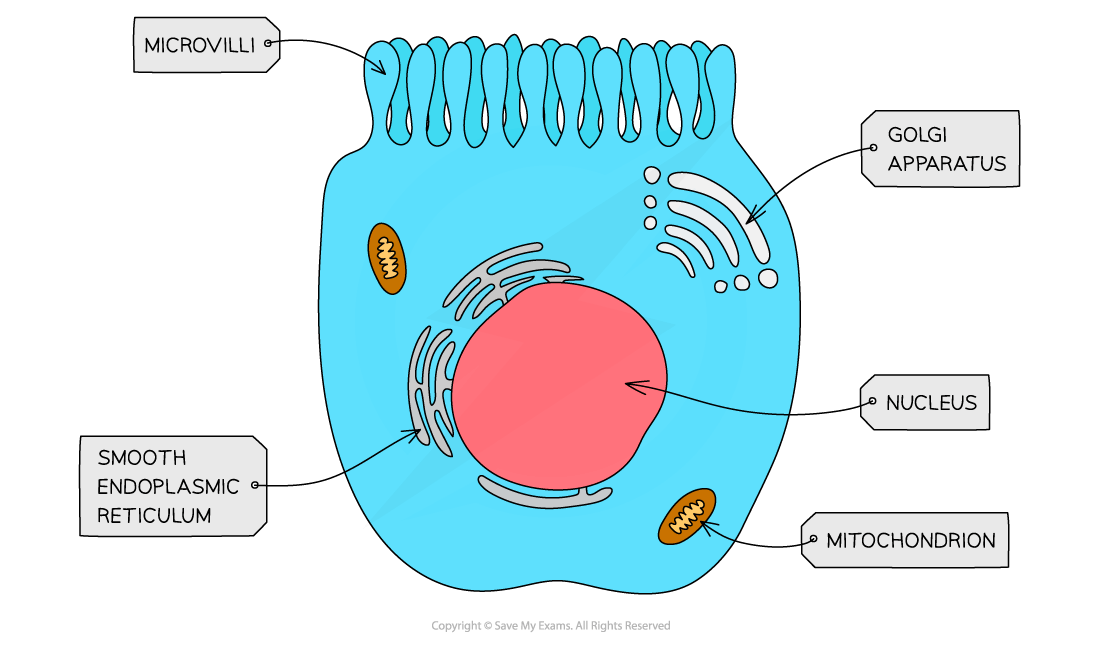
Microvilli are cell membrane projections that increase the surface area for absorption
Microvilli are found in parts of the body that carry out absorption, e.g.
The lining of the small intestine
The kidney tubules
Examiner Tips and Tricks
Be careful not to confuse microvilli with villi. Villi are much larger structures made up of several layers of cells, while microvilli are found on the surfaces of individual cells. Microvilli will be present on the outermost layer of cells that make up the villi!
Cell wall
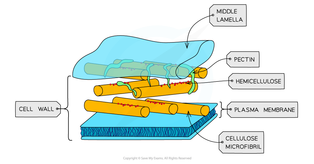
Cell walls are outside cell surface membranes and offer structural support to some types of cell
Structural support is provided by the polysaccharide cellulose in plants, and by chitin in fungi
Cell walls are freely permeable and do not play a role in controlling the movement of substances into and out of cells
Chloroplasts
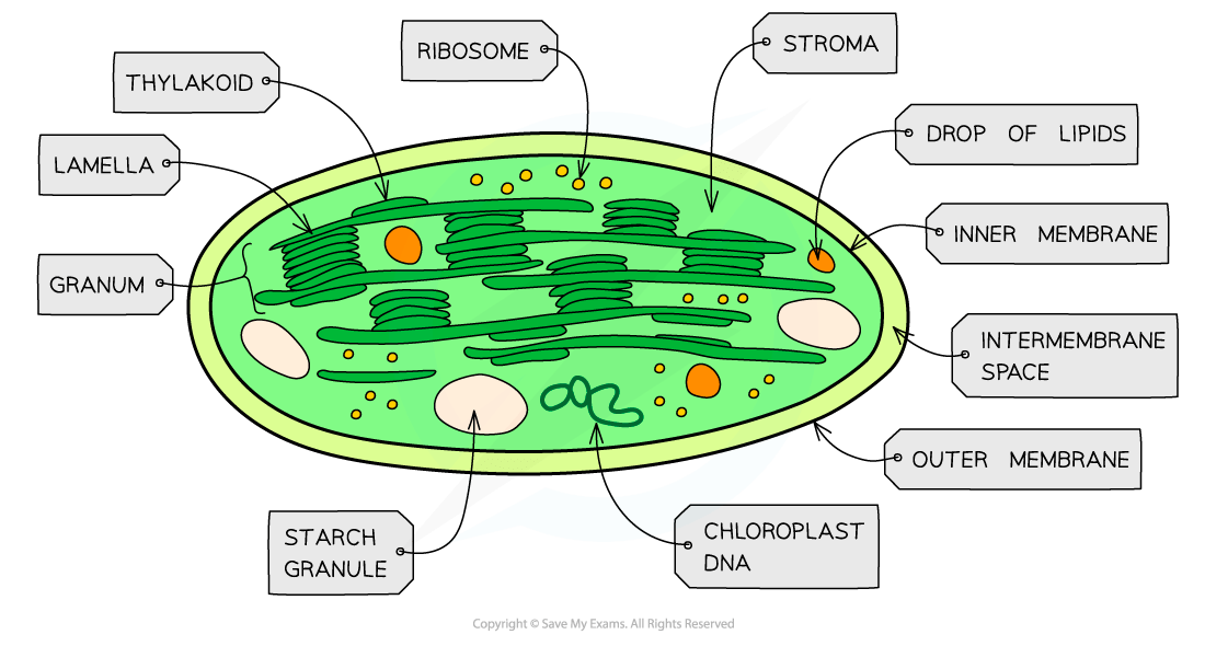
Chloroplasts are larger than mitochondria, and are also surrounded by a double-membrane
Membrane-bound compartments called thylakoids stack together to form structures called grana
Grana are joined together by lamellae
Photosynthetic pigments such as chlorophyll are found in the membranes of the thylakoids, where their role is to absorb light energy for photosynthesis
Chloroplasts contain small circular pieces of DNA and ribosomes used to synthesise proteins needed in chloroplast replication and photosynthesis
Plasmodesmata
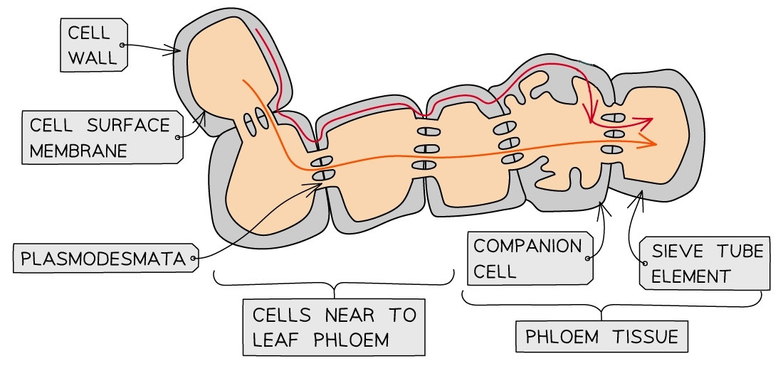
Plasmodesmata are bridges of cytoplasm between neighbouring plant cells
They allow the transfer of substances between plant cells
Large permanent vacuole
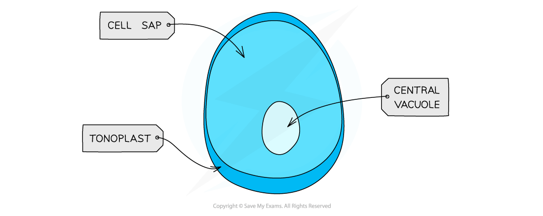
Large permanent vacuoles are found in plant cells, where they store cell sap and provide additional structural support to cells
Vacuoles are sometimes found in animal cells, but these will be small and temporary
Vacuoles are surrounded by the tonoplast, which is a partially permeable membrane

Unlock more, it's free!
Did this page help you?