Doppler Effect in Ultrasound (OCR A Level Physics): Revision Note
Exam code: H556
Doppler Effect in Ultrasound
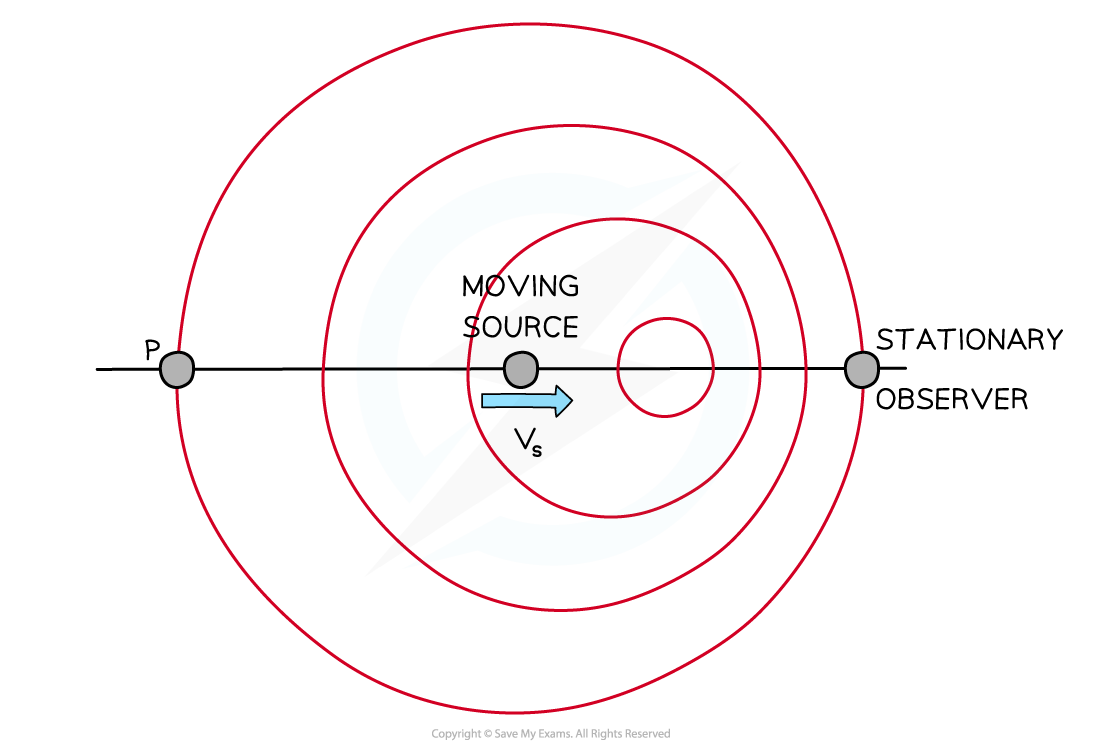
The Doppler effect is defined as:
The frequency change of a wave due to the relative motion between a source and an observer
When the source starts to move towards the observer, the wavelength of the waves is shortened
The sound therefore appears at a higher frequency to the observer
The frequency is increased when the source is moving towards the observer
The frequency is decreased when the source is moving away from the observer
This applies to ultrasound in the exact same way as it does to sound waves

Doppler effect of a moving source and stationary observer
For a wave of frequency, f0, that reflects at a moving boundary, the new frequency, fr, of the reflected pulse is given by:
Where:
f0 = frequency of original wave (Hz)
fr = frequency of reflected wave (Hz)
v = velocity of the moving boundary (m s−1)
c = velocity of the wave (m s−1)
The factor of 2 appears due to the wave travelling to the boundary and back again
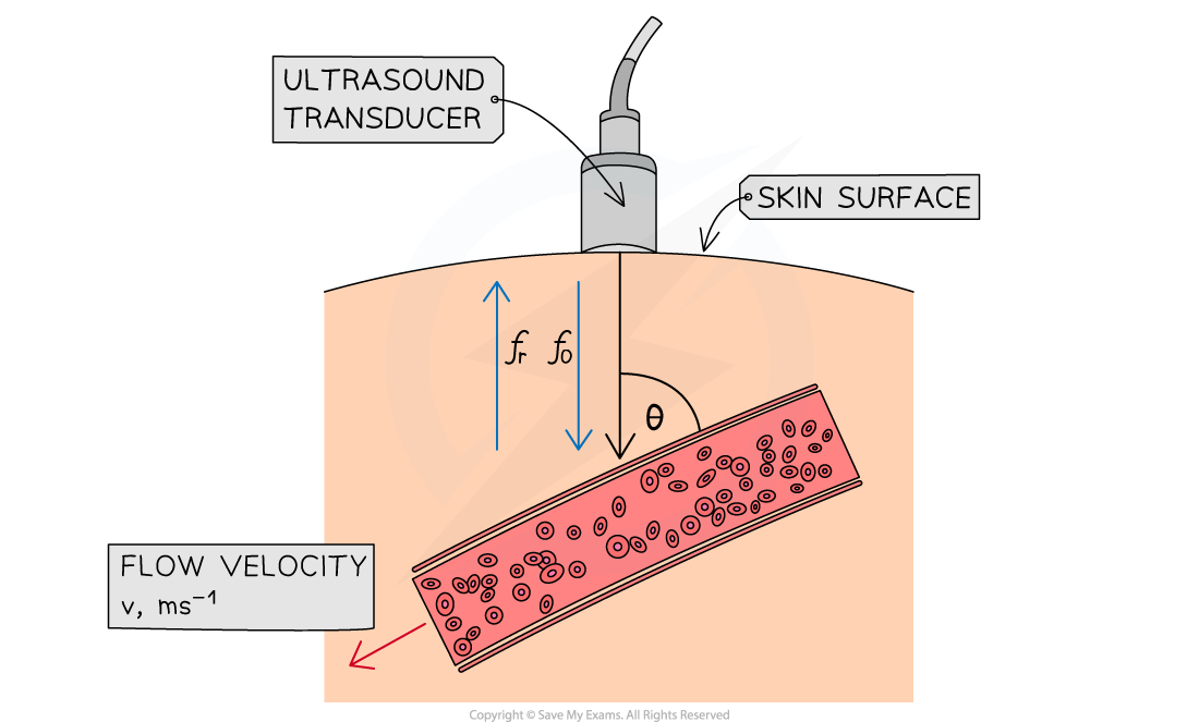
In medicine, Doppler imaging can be used as a non-invasive technique to measure the speed of blood flow in the heart or in an artery
This is effective because blood contains iron which is very reflective
The journey of the ultrasound is as follows:
Pulses of ultrasound are emitted from a transducer into a blood vessel
The ultrasound pulses are reflected by moving blood cells
The moving blood causes a shift in the frequency of the ultrasound
The transducer detects an increase in frequency if the blood is moving towards the transducer and a decrease if it is moving away

This frequency shift, Δf, can be calculated using the equation:
Where:
f0 = frequency of ultrasound emitted by the transducer (Hz)
fr = frequency of ultrasound received by the transducer (Hz)
Δf = difference between emitted and received frequencies (Hz)
v = velocity of the blood (m s−1)
c = velocity of ultrasound in blood (m s−1)
θ = angle between the transducer and the blood vessel (°)
This can be rearranged to give:
Worked Example
A patient is undergoing diagnostic tests to determine the cause of an underlying heart condition. Blood flow tests are done on the patient's aorta using ultrasound imaging.
Ultrasound of frequency 8.5 MHz is transmitted from a transducer through the skin into the aorta. The reflected frequency is found to be 8.495 MHz. The angle the transducer makes with the skin is 15°.
The velocity of sound in blood is 1570 m s−1 and the aorta has an internal diameter of 30 mm.
Calculate:
a) The speed of the blood flow in the aorta.
b) The volume of blood, in cm3, moving past any point in 1 s.
Answer:
Part (a)
Step 1: List the known quantities
Transmitted frequency of the ultrasound, f0 = 8.5 MHz
Reflected frequency of the ultrasound, fr = 8.495 MHz
Change in frequency, Δf = 8.5 − 8.495 = 0.005 MHz
Velocity of sound in blood, c = 1570 m s−1
Angle between the transducer and skin, θ = 15°
Step 2: Write out the equation for Doppler shift of ultrasound
Step 3: Rearrange for the speed, v, and calculate
= 0.478
v = 0.48 m s−1
Part (b)
Step 1: List the known quantities
Radius of the aorta, r = 15 mm = 1.5 cm
Blood flow rate, v = 0.48 m s−1 = 48 cm s−1
Step 2: Calculate the cross-sectional area of the aorta
Cross-sectional area = πr2 = π(1.5)2 = 7.1 cm2
Step 3: Determine the volume of blood passing through the aorta each second
Volume flow rate = cross-sectional area (cm2) × blood flow rate (cm s−1)
Volume flow rate = 7.1 × 48 = 341 cm3 s−1
Step 4: State the volume of blood in 1 s
The volume of blood that flows in 1 s = 341 cm3

Unlock more, it's free!
Did this page help you?