Detecting Gamma-Rays from PET Scanning (Cambridge (CIE) A Level Physics): Revision Note
Exam code: 9702
Calculating energy of gamma-ray photons
In the annihilation process, mass, energy and momentum are conserved
The gamma-ray photons have an energy and frequency that is determined solely by the mass-energy of the positron-electron pair
The energy of a photon is given by:
The momentum of a photon is given by
Where:
me = mass of the electron or positron (kg)
h = Planck's constant (J s)
f = frequency of the photon (Hz)
c = the speed of light in a vacuum (m s–1)
Worked Example
Fluorine-18 decays by β+ emission. The positron emitted collides with an electron and annihilates producing two γ-rays.
(a) Calculate the energy released when a positron and an electron annihilate.
(b) Calculate the frequency of the γ-rays emitted.
(c) Calculate the momentum of one of the γ-rays.
Answer:
Part (a)
Step 1: Write down the known quantities
Mass of an electron = mass of a positron, me = 9.11 × 10⁻³¹ kg
Total mass is equal to the mass of the electron and positron = 2me
Step 2: Write out the equation for mass-energy equivalence
Step 3: Substitute in values and calculate energy
Part (b)
Step 1: Determine the energy of one photon
Planck's constant, h = 6.63 × 10⁻³⁴ J s
Two photons are produced, so, the energy of one photon is equal to half of the total energy from part (a):
Step 2: Write out the equation for the energy of a photon
Step 3: Rearrange for frequency and calculate
Part (c)
Step 1: Write out the equation for the momentum of a photon
Step 2: Substitute in values and calculate momentum
Detecting gamma-rays from PET scanning
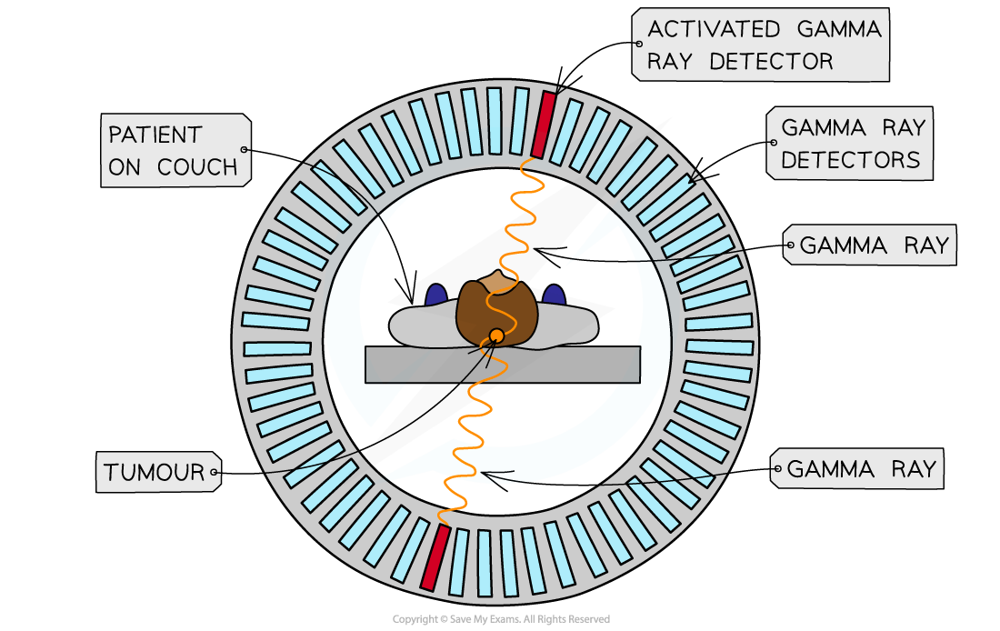
The patient lays stationary in a tube surrounded by a ring of detectors
Images of slices of the body can be taken to show the position of the radioactive tracers
The detector consists of two parts:
A crystal scintillator
A photomultiplier
Crystal scintillator
When the gamma-ray (γ-ray) photon is incident on a crystal, an electron in the crystal is excited to a higher energy state
As the excited electron travels through the crystal, it excites more electrons
When the excited electrons move back down to their original state, the lost energy is transmitted as visible light photons
Photomultiplier
The photons produced by the scintillator are very faint
Therefore, they need to be amplified and converted to an electrical signal by a photomultiplier tube
PET scanner

Detecting gamma rays with a PET scanner
Creating an image from PET scanning
The γ rays travel in straight lines in opposite directions when formed from a positron-electron annihilation
This is due to the conservation of momentum
They hit the detectors in a line – known as the line of response
The tracers will emit lots of γ rays simultaneously, and the computers will use this information to create an image
The more photons formed at a particular point, the more tracer that is present in the tissue being studied, and this will appear as a bright point on the image
An image of the tracer concentration in the tissue can be created by processing the arrival times of the gamma-ray photons

Unlock more, it's free!
Did this page help you?