The PET Scanner (AQA A Level Physics): Revision Note
Exam code: 7408
Positron Emission Tomography
Positron Emission Tomography (PET) is defined as:
A type of nuclear medical procedure that images tissues and organs by measuring the metabolic activity of the cells of body tissues
In PET scanning, a beta-plus emitting radioactive tracer is used in order to stimulate positron-electron annihilation to produce gamma photons
These are then detected using a ring of gamma cameras
Principles of PET Scanning
Before the scan
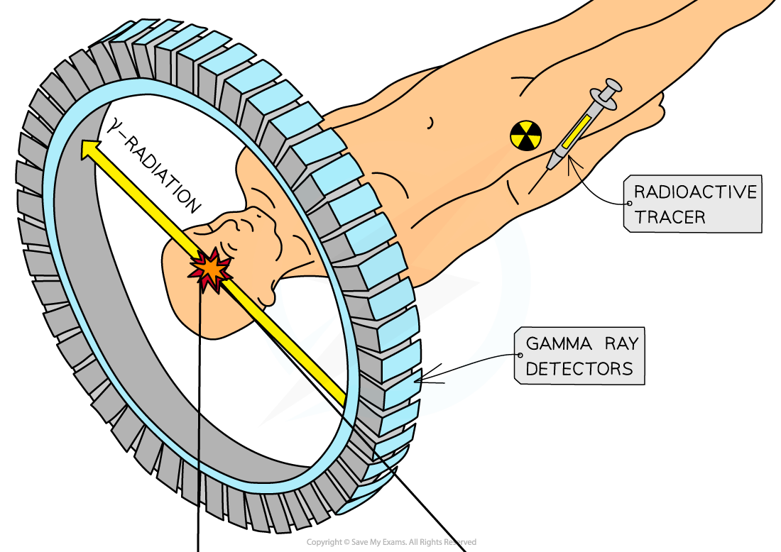
The patient is injected with a beta-plus emitting isotope, usually fluorine-18 (F-18)
During the scan
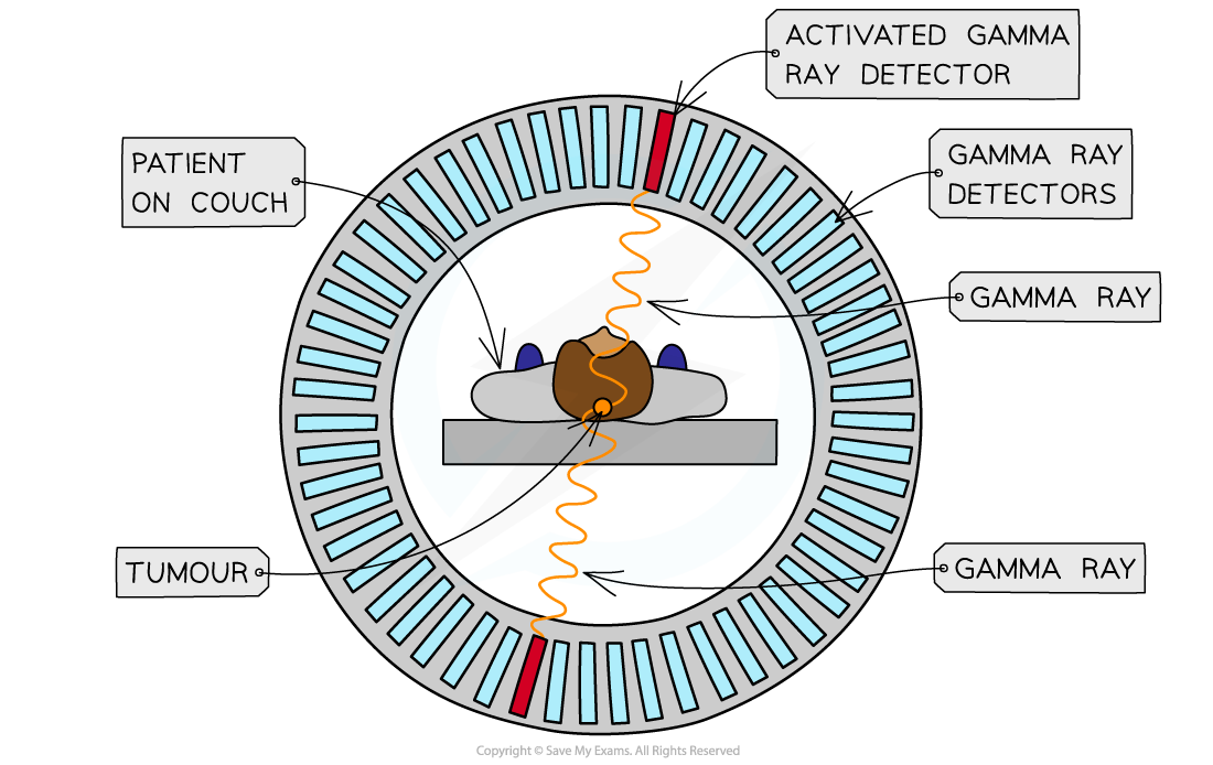
The part of the body being studied is surrounded by a ring of gamma cameras
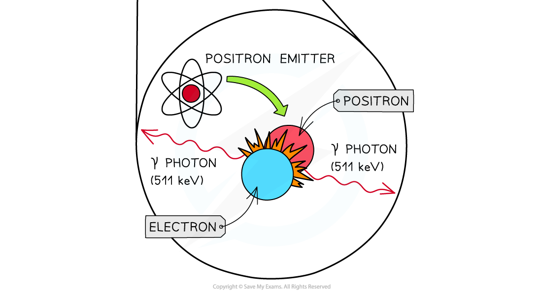
The positrons from the F-18 nuclei annihilate with electrons in the patient
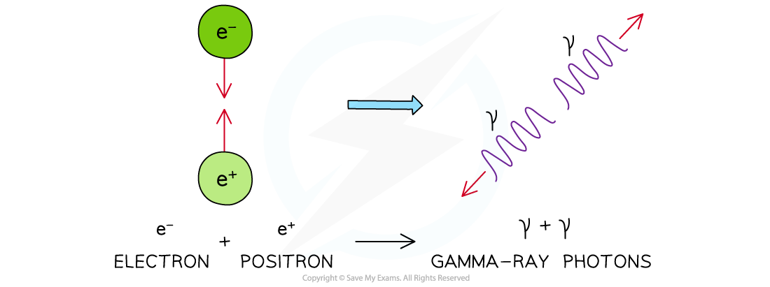
The annihilation of a positron and an electron produces two identical gamma photons travelling in opposite directions
The delay time between these two gamma-ray photons is used to determine the location of the annihilation due to the F-18 tracer
Photons that do not arrive within a nanosecond of each other are ignored since they cannot have come from the same point
After the scan
The signals from the gamma camera detectors are sent to a computer which builds up an image
A PET Scanner

Detecting gamma rays with a PET scanner
Annihilation
Annihilation can happen between any particle and antimatter counterpart, but in PET scanning, only the electron and positron interaction is considered
When a positron is emitted from a tracer in the body, it travels less than a millimetre before annihilating with an atomic electron
As with all collisions, mass, energy and momentum are always conserved
An Electron-Positron Annihilation

Annihilation of a positron and electron to form two gamma-ray photons
The gamma-ray photons produced each have an energy of 512 keV
This is the same each time as determined by the mass-energy equivalence of the positron-electron pair
The energy of each photon is given by
Where:
= mass of the electron or positron (kg)
= Planck's constant (J s)
= frequency of the photon (Hz)
= the speed of light in a vacuum (m s–1)
Diagnosis Using PET Scanning
The signals produced by the photomultiplier tubes are used to produce an image
The γ rays travel in straight lines in opposite directions when formed from the annihilation of the positron and electron
This happens in order to conserve momentum
They hit the detectors in a line – known as the line of response
The tracers will emit lots of γ rays simultaneously, and the computers will use this information to create an image
The more photons from a particular point, the more tracer that is present in the tissue being studied, and this will appear as a bright point on the image
An image of the tracer concentration in the tissue can be created by processing the arrival times of the gamma-ray photons
Annihilation in the PET Scanner


Annihilation of a positron and an electron is the basis of PET Scanning
Once the tracer is introduced to the body it has a short half-life, so, it begins emitting positrons
immediately
This allows for a short exposure time to the radiation
A short half-life does mean the patient needs to be scanned quickly and not all hospitals have access to expensive PET scanners
Worked Example
Fluorine-18 decays by β+ emission. The positron emitted collides with an electron and annihilates producing two γ-rays.
(a) Calculate the total energy released when a positron and an electron annihilate.
(b) Show that each gamma ray has an energy of 512 keV.
(c) Calculate the frequency of the gamma rays.
Answer:
Part (a)
Step 1: Write down the known quantities
Mass of an electron = mass of a positron,
= 9.11 × 10⁻³¹ kg
Total mass = mass of the electron and positron =
Speed of light,
= 3.00 × 10⁸ m s–1
Step 2: Write out the equation for mass-energy equivalence
Step 3: Substitute in values and calculate energy E
Part (b)
Step 1: Determine the energy of one photon
Planck's constant, h = 6.63 × 10⁻³⁴ J s
Two photons are produced, so, the energy of one photon is equal to half of the total energy from part (a):
Note: if you keep the exact value for energy in your calculator, you would have E = 8.199 × 10⁻¹⁴ J
Step 2: Convert the energy in J into eV:
1 eV = 1.6 × 10⁻¹⁹ J
Part (c)
Step 1: Write out the equation for the energy of a photon
Step 2: Rearrange for frequency f and calculate

Unlock more, it's free!
Did this page help you?