Microarrays (Cambridge (CIE) A Level Biology): Revision Note
Exam code: 9700
Microarrays
Microarrays are laboratory tools used to detect the expression of thousands of genes at the same time and to identify the genes present in an organism’s genome
Microarrays are used in different ways, e.g.
Pathology: comparison between healthy cells and diseased cells to find the characteristics of the disease
Biotechnology (e.g. in agriculture to identify insect pests)
Crime (e.g. forensic analysis)
As large numbers of genes can be studied in a short period of time, microarrays have been very valuable to scientists
The microarray consists of a small (usually 2cm2) piece of glass, plastic or silicon (also known as chips) that have probes attached to a spot (called a gene spot) in a grid pattern
There can be 10 000 or more spots per cm2
Probes are short lengths of single-stranded DNA (oligonucleotides) or RNA which are synthesised to be complementary for a specific base sequence (this sequence depends on the purpose of the microarray)
Using microarrays to analyse genomes
DNA is collected from the species going to be compared
Restriction enzymes are used to cut the DNA into fragments
These fragments are denatured to create single-stranded DNA molecules
These DNA fragments are labelled using fluorescent tags (the fragments from the different sources are tagged in different colours, usually red and green)
Once these fragments are mixed together they are allowed to hybridise with the probes on the microarray
After a set period of time, any DNA that did not hybridise with the probes is washed off
The microarray is then examined using ultraviolet light (which causes the tags to fluoresce) or scanned (colours are detected by the computer and the information is analysed and stored)
The presence of the colour indicates where hybridisation has occurred, as the DNA fragment is complementary to the probe
If red and green fluorescent spots appear then only one species of DNA has hybridised, however, if the spot is yellow then both species have hybridised with that DNA fragment, which suggests that both species have that gene in common
If a spot lacks colour that indicates the gene is not present in either species
Using microarrays to study gene expression
When genes are being expressed or are in their active state, many copies of mRNA are produced by transcription
The corresponding proteins are then produced from these mRNAs during translation
Thus scientists can indirectly, by assessing the quantity of mRNAs, determine which genes are being expressed in the cells
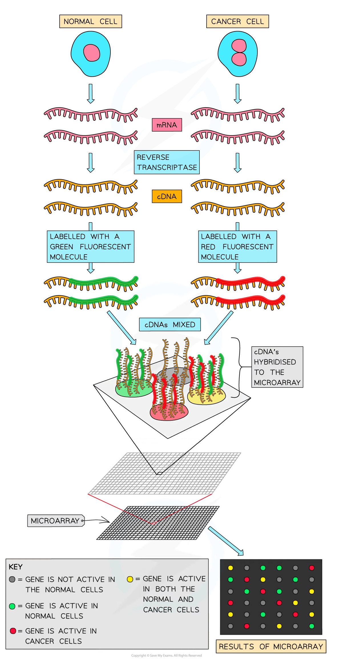
Microarrays can be used to detect whether a gene is being expressed (a method used to research cancerous vs non-cancerous cells) by detecting the quantity of mRNA present
To compare which genes are being expressed using microarrays the following steps occur:
mRNA is collected from both types of cells and reverse transcriptase is used to convert mRNA into cDNA
PCR may be used to increase the quantity of cDNA (this occurs for all samples to remain proportional so a comparison can be made when analysis occurs)
Fluorescent tags are added to the cDNA
The cDNA is then denatured to produce single-stranded DNA
The single-stranded DNA molecules are allowed to hybridise with the probes on the microarray
When the ultraviolet light is shone on the microarray the spots that fluoresce indicate that the gene was transcribed (expressed) and the intensity of the light emitting from the spots indicates the quantity of mRNA produced (i.e. how active the gene is)
If the light being emitted is of high intensity then many mRNA were present, while a low intensity emission indicates few mRNA are present

Examiner Tips and Tricks
The colours of the fluorescent tags (red, green and yellow) indicate whether a gene is present whereas the intensity of light emitted indicates the level of gene expression (the more light, the more the gene was being expressed) and this relates to the quantity of mRNA present.

Unlock more, it's free!
Was this revision note helpful?
