Resolution & Magnification (Cambridge (CIE) A Level Biology): Revision Note
Exam code: 9700
Resolution & magnification
Magnification
Magnification is the number of times that a real-life specimen has been enlarged to give a larger view/image
E.g. a magnification of x100 means that a specimen has been enlarged 100 times to give the image shown
A light microscope has two types of lens which allow it to achieve different levels of magnification:
An eyepiece lens, which often has a magnification of x10
A series of objective lenses, each with a different magnification, e.g. x4, x10, x40 and x100
To calculate the total magnification of a specimen being viewed, the magnification of the eyepiece lens and the objective lens are multiplied together:
Total magnification = eyepiece lens magnification x objective lens magnification
Resolution
The resolution of a microscope is its ability to distinguish two separate points on an image as separate objects; this determines the ability of a microscope to show detail
If resolution is too low then two separate objects will be observed as one point, and an image will appear blurry, or an object will not be visible at all
The resolution of a microscope limits the magnification that it can usefully achieve; there is no point in increasing the magnification to a higher level if the resolution is poor
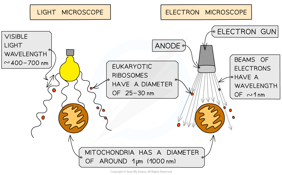
The resolution of a light microscope is limited by the wavelength of light
Visible light falls within a set range of light wavelengths; 400-700 nm
The resolution of a light microscope cannot be smaller than half the wavelength of visible light
The shortest wavelength of visible light is 400 nm, so the maximum resolution of a light microscope is 200 nm
E.g. the structure of a phospholipid bilayer cannot be observed under a light microscope due to low resolution:
The width of the phospholipid bilayer is about 10 nm
The maximum resolution of a light microscope is 200 nm, so any points that are separated by a distance of less than 200 nm, such as the 10 nm phospholipid bilayer, cannot be resolved by a light microscope and therefore will not be distinguishable as separate objects
Electron microscopes have a much higher resolution, and therefore magnification, than light microscopes as electrons have a much smaller wavelength than visible light
Electron microscopes can achieve a resolution of 0.5 nm

Examiner Tips and Tricks
Don’t assume that increasing magnification will let you see more detail — it won’t if the resolution is too low.
Remember: magnification makes things bigger, resolution makes things clearer.

Unlock more, it's free!
Did this page help you?